Correctly label the bones in the wrist – Understanding the intricacies of the wrist bones is essential for accurate anatomical labeling. This guide provides a comprehensive overview of the structure, function, and labeling techniques of the wrist bones, empowering medical professionals, students, and researchers with the knowledge to confidently and precisely identify these vital structures.
Anatomical Overview

The wrist is a complex joint that connects the forearm to the hand. It is made up of eight carpal bones, the distal ends of the radius and ulna, and a network of ligaments. The wrist bones are arranged in two rows, with the proximal row articulating with the radius and ulna and the distal row articulating with the metacarpals of the hand.
The wrist joint allows for a wide range of motion, including flexion, extension, radial and ulnar deviation, and pronation and supination. The carpal bones are responsible for transmitting forces between the forearm and the hand, and they also provide stability to the wrist joint.
Carpal Bones

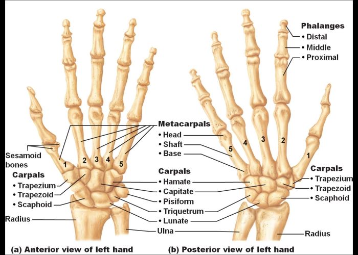
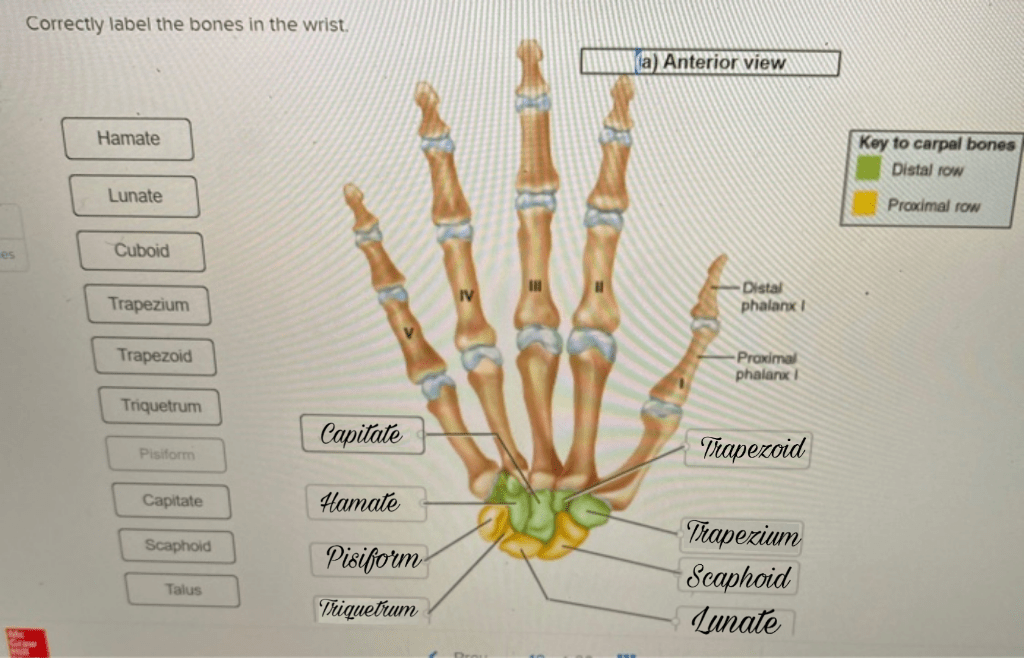
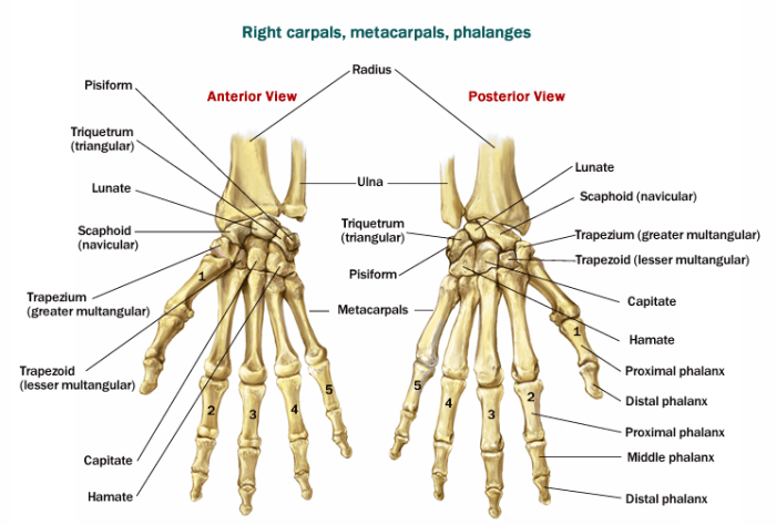
The eight carpal bones are divided into two rows, with four bones in each row. The proximal row, from lateral to medial, consists of the scaphoid, lunate, triquetrum, and pisiform bones. The distal row, from lateral to medial, consists of the trapezium, trapezoid, capitate, and hamate bones.
Each carpal bone has a unique shape and function. The scaphoid bone is the largest of the carpal bones and it articulates with the radius, lunate, capitate, and trapezium bones. The lunate bone is saddle-shaped and it articulates with the radius, scaphoid, capitate, and hamate bones.
The triquetrum bone is triangular in shape and it articulates with the ulna, lunate, hamate, and pisiform bones. The pisiform bone is a small, pea-shaped bone that articulates with the triquetrum bone.
The trapezium bone is the most lateral bone in the distal row and it articulates with the scaphoid, trapezoid, and first metacarpal bones. The trapezoid bone is located between the trapezium and capitate bones and it articulates with the scaphoid, trapezium, and second metacarpal bones.
The capitate bone is the largest bone in the distal row and it articulates with the scaphoid, lunate, hamate, and second and third metacarpal bones. The hamate bone is the most medial bone in the distal row and it articulates with the triquetrum, lunate, capitate, and fourth and fifth metacarpal bones.
Distal Radius and Ulna
The distal radius and ulna are the two forearm bones that articulate with the carpal bones. The distal radius is a triangular bone that forms the lateral aspect of the wrist joint. The distal ulna is a smaller, hook-shaped bone that forms the medial aspect of the wrist joint.
The distal radius and ulna play an important role in wrist articulation. The distal radius articulates with the scaphoid, lunate, and triquetrum bones, while the distal ulna articulates with the triquetrum bone. The distal radius and ulna also contribute to wrist stability by forming a strong bony arch that supports the carpal bones.
Wrist Ligaments

The wrist joint is stabilized by a network of ligaments. The major ligaments of the wrist include the:
- Dorsal carpal ligament:This ligament runs across the back of the wrist and connects the carpal bones to the radius and ulna.
- Palmar carpal ligament:This ligament runs across the front of the wrist and connects the carpal bones to the metacarpals.
- Scapholunate ligament:This ligament connects the scaphoid and lunate bones and prevents them from dislocating.
- Lunotriquetral ligament:This ligament connects the lunate and triquetrum bones and prevents them from dislocating.
- Triquetrohamate ligament:This ligament connects the triquetrum and hamate bones and prevents them from dislocating.
- Pisotriquetral ligament:This ligament connects the pisiform and triquetrum bones and prevents the pisiform bone from dislocating.
Clinical Significance: Correctly Label The Bones In The Wrist

Misalignment or injury to the wrist bones can have a significant impact on wrist function. Common wrist disorders include:
- Carpal tunnel syndrome:This is a condition in which the median nerve is compressed in the carpal tunnel, which is a narrow passageway in the wrist. Carpal tunnel syndrome can cause pain, numbness, and tingling in the hand and fingers.
- Tendonitis:This is a condition in which the tendons that connect the muscles to the bones in the wrist become inflamed. Tendonitis can cause pain, swelling, and stiffness in the wrist.
- Arthritis:This is a condition in which the cartilage that lines the wrist joint becomes damaged. Arthritis can cause pain, swelling, and stiffness in the wrist.
- Wrist fractures:These are breaks in the bones of the wrist. Wrist fractures can be caused by falls, sports injuries, or other accidents.
Essential Questionnaire
What is the most important anatomical landmark for labeling wrist bones?
The styloid process of the radius is a prominent landmark that aids in identifying and labeling the carpal bones.
How can I avoid common errors when labeling wrist bones?
Pay attention to the shape, size, and orientation of each bone, and refer to anatomical models or images for guidance.